
- Current
- Browse
- Collections
-
For contributors
- For Authors
- Instructions to authors
- Article processing charge
- e-submission
- For Reviewers
- Instructions for reviewers
- How to become a reviewer
- Best reviewers
- For Readers
- Readership
- Subscription
- Permission guidelines
- About
- Editorial policy
Search
- Page Path
- HOME > Search
Original Article
- Basic Research
- Hyperglycemia-Suppressed SMARCA5 Disrupts Transcriptional Homeostasis to Facilitate Endothelial Dysfunction in Diabetes
- Ju Wang, Hui Zhou, Jinhua Shao, Shu Zhang, Jing Jin
- Diabetes Metab J. 2023;47(3):366-381. Published online March 6, 2023
- DOI: https://doi.org/10.4093/dmj.2022.0179
- 1,713 View
- 98 Download
-
 Abstract
 PDF
PDF Supplementary Material
 PubReader
  ePub
ePub - Background
Dysfunction of vascular endothelial cells (ECs) plays a central role in the pathogenesis of cardiovascular complications in diabetes. SWI/SNF-related matrix-associated actin-dependent regulator of chromatin subfamily A member 5 (SMARCA5) is a key regulator of chromatin structure and DNA repair, but its role in ECs remains surprisingly unexplored. The current study was designed to elucidate the regulated expression and function of SMARCA5 in diabetic ECs.
Methods
SMARCA5 expression was evaluated in ECs from diabetic mouse and human circulating CD34+ cells using quantitative reverse transcription polymerase chain reaction and Western blot. Effects of SMARCA5 manipulation on ECs function were evaluated using cell migration, in vitro tube formation and in vivo wound healing assays. Interaction among oxidative stress, SMARCA5 and transcriptional reprogramming was elucidated using luciferase reporter assay, electrophoretic mobility shift assay and chromatin immunoprecipitation.
Results
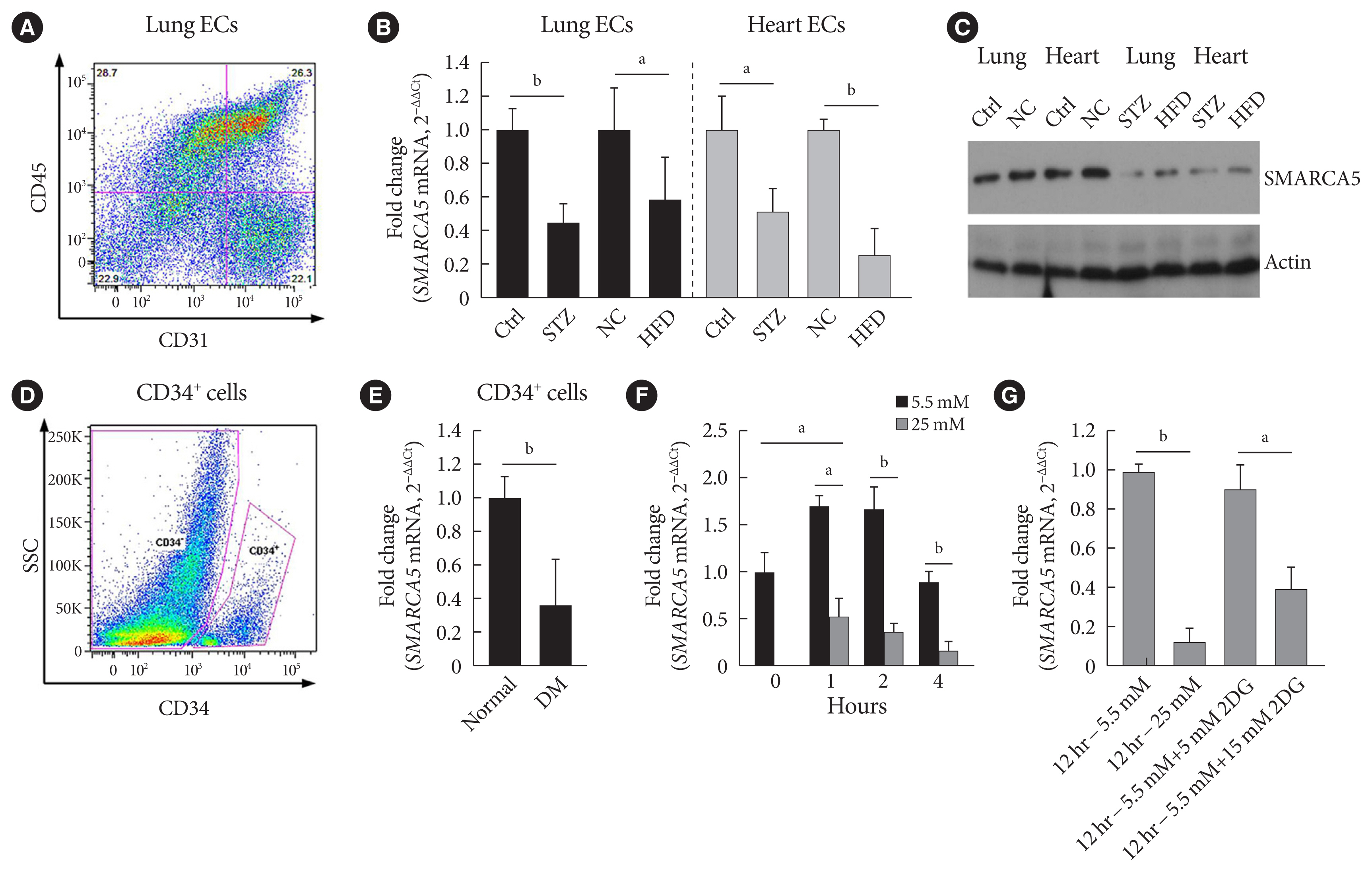
Endothelial SMARCA5 expression was significantly decreased in diabetic rodents and humans. Hyperglycemia-suppressed SMARCA5 impaired EC migration and tube formation in vitro, and blunted vasculogenesis in vivo. Contrarily, overexpression of SMARCA5 in situ by a SMARCA5 adenovirus-incorporated hydrogel effectively promoted the rate of wound healing in a dorsal skin punch injury model of diabetic mice. Mechanistically, hyperglycemia-elicited oxidative stress suppressed SMARCA5 transactivation in a signal transducer and activator of transcription 3 (STAT3)-dependent manner. Moreover, SMARCA5 maintained transcriptional homeostasis of several pro-angiogenic factors through both direct and indirect chromatin-remodeling mechanisms. In contrast, depletion of SMARCA5 disrupted transcriptional homeostasis to render ECs unresponsive to established angiogenic factors, which ultimately resulted in endothelial dysfunction in diabetes.
Conclusion
Suppression of endothelial SMARCA5 contributes to, at least in part, multiple aspects of endothelial dysfunction, which may thereby exacerbate cardiovascular complications in diabetes.
Review
- Impaired Wound Healing in Diabetes Mellitus.
- Min Jeong Kwon, Jeong Hyun Park
- Korean Diabetes J. 2009;33(2):83-90. Published online April 1, 2009
- DOI: https://doi.org/10.4093/kdj.2009.33.2.83
- 1,805 View
- 36 Download
- 3 Crossref
-
Abstract
PDF
- The normal healing of a cutaneous wound is achieved via well-orchestrated integration of complex biological and molecular events of cell migration, proliferation, extracellular matrix deposition and tissue remodeling. Chronic wounds fail to progress through the normal stages of healing, and enter a state of pathologic inflammation. Complicated diabetic patients show delayed wound healing caused by multiple factors including vascular insufficiency, abnormalities of the biochemical environment and hyperglycemia per se. Novel technologies including growth factor therapy, gene therapy, stem cell technologies, synthetic skins and hyperbaric oxygen treatment are under development. In the near future, these therapeutic strategies will be clinically available.
-
Citations
Citations to this article as recorded by
- Promotion of wound healing through low-fluence ablative fractional laser treatment in diabetic mice
Han Na Lee, Jung Min Bae, Bon Cheol Leo Goo, Young Min Park
Lasers in Medical Science.2019; 34(2): 421. CrossRef - Ethanol Extracts of Chungkookjang Stimulate the Proliferation and Migration of Human Umbilical Vascular Endothelial Cells
Jae Sung Hwang, Dae Il Sung, Whan Myung Lee, Young Shin Chung, Han Bok Kim
The Korean Journal of Microbiology.2014; 50(3): 223. CrossRef - Comparison of Outcome of Trabeculectomy With Mitomycin C and Ahmed Valve Implantation for Uveitic Glaucoma
Joo Yeon Kim, Hyoung Sub Shim, Hwang Ki Kim, Yong Ho Sohn
Journal of the Korean Ophthalmological Society.2010; 51(4): 575. CrossRef
- Promotion of wound healing through low-fluence ablative fractional laser treatment in diabetic mice
Original Article
- Cloning of Novel Epidermal Growth Factor (EGF) Plasmid for Gene Therapy on Diabetic Foot Ulcer.
- Hye Sook Chung, Chang Shin Yoon, Min Jeong Kwon, Mi Kyung Kim, Soon Hee Lee, Kyung Soo Ko, Byung Doo Rhee, Jeong Hyun Park
- Korean Diabetes J. 2008;32(2):131-140. Published online April 1, 2008
- DOI: https://doi.org/10.4093/kdj.2008.32.2.131
- 1,806 View
- 38 Download
- 1 Crossref
-
Abstract
PDF
- BACKGROUND
Epidermal Growth Factor (EGF) is one of the important growth factors involved in the epithelialization during cutaneous wound healing. Peptide EGF has been used for the treatment of diabetic foot ulcer. But the inferiority of cost-effectiveness and the inconvenience of daily application might have restricted its wide clinical usage. EGF gene therapy could dramatically improve the efficacy and inconvenience through long-term expression and bypassing the EGF degradation by hostile non-specific proteinases expressed in the wound bed. METHODS: EGF DNAs were amplified via PCR. For the more effective secretion from the transfected cell, we inserted furin cleavage site into EGF plasmids. The efficacy of novel plasmid pbeta-EGF was verified by transfection into the various animal cell lines, and the biologic potency of expressed EGF was confirmed via phosphorylation of PI3K and GSK3beta by Western blotting. RESULTS: We tested various kinds of human EGFs. One of the human EGF isoforms, EGF(828) including a membrane-anchoring domain was successfully released as the mature EGF protein in the cell culture media. Also EGF plasmid including furin cleavage site showed more than 2-fold increased EGF expression compared with the sequence without furin cleavage site. CONCLUSION: In conclusion, these findings suggest that mature EGF could be released easily out of cells by modifying EGF DNA sequence. Our novel EGF plasmid DNA could markedly increase the efficiency of non-viral gene therapy for diabetic foot ulcer. -
Citations
Citations to this article as recorded by- Effective healing of diabetic skin wounds by using nonviral gene therapy based on minicircle vascular endothelial growth factor DNA and a cationic dendrimer
Min J. Kwon, Songhie An, Sunghyun Choi, Kihoon Nam, Hye S. Jung, Chang S. Yoon, Jung H. Ko, Hye J. Jun, Tae K. Kim, Soo J. Jung, Jeong H. Park, Yan Lee, Jong‐Sang Park
The Journal of Gene Medicine.2012; 14(4): 272. CrossRef
- Effective healing of diabetic skin wounds by using nonviral gene therapy based on minicircle vascular endothelial growth factor DNA and a cationic dendrimer
 First
First Prev
Prev


