An In Vitro Model to Probe the Regulation of Adipocyte Differentiation under Hyperglycemia
Article information
Abstract
Background
The aim of this study was an in vitro investigation of the effect of high glucose concentration on adipogenesis, as prolonged hyperglycemia alters adipocyte differentiation.
Methods
3T3-L1 preadipocytes differentiated in the presence of varying concentrations of glucose (25, 45, 65, 85, and 105 mM) were assessed for adipogenesis using AdipoRed (Lonza) assay. Cell viability and proliferation were measured using MTT reduction and [3H] thymidine incorporation assay. The extent of glucose uptake and glycogen synthesis were measured using radiolabelled 2-deoxy-D-[1-3H] glucose and [14C]-UDP-glucose. The gene level expression was evaluated using reverse transcription-polymerase chain reaction and protein expression was studied using Western blot analysis.
Results
Glucose at 105 mM concentration was observed to inhibit adipogenesis through inhibition of CCAAT-enhancer-binding proteins, sterol regulatory element-binding protein, peroxisome proliferator-activated receptor and adiponectin. High concentration of glucose induced stress by increasing levels of toll-like receptor 4, nuclear factor κB and tumor necrosis factor α thereby generating activated preadipocytes. These cells entered the state of hyperplasia through inhibition of p27 and proliferation was found to increase through activation of protein kinase B via phosphoinositide 3 kinase dependent pathway. This condition inhibited insulin signaling through decrease in insulin receptor β. Although the glucose transporter 4 (GLUT4) protein remained unaltered with the glycogen synthesis inhibited, the cells were found to exhibit an increase in glucose uptake via GLUT1.
Conclusion
Adipogenesis in the presence of 105 mM glucose leads to an uncontrolled proliferation of activated preadipocytes providing an insight towards understanding obesity.
INTRODUCTION
The state of high glucose has been known to induce the process of adipogenesis [1,2]. Prolonged condition of hyperglycemia alters the production of adipocytokines and induces cytokine mediated stress and inflammation [3]. Increased levels of tumor necrosis factor α (TNFα) and toll-like receptor 4 (TLR4) during inflammation have been found to inhibit differentiation of preadipocytes into functional adipocytes [4,5]. These undifferentiated preadipocytes, also called "activated" preadipocytes, account for an increased release of chemokines and cytokines from the adipose tissue under obese conditions [6]. The present study attempts to understand the signaling events regulating preadipocyte differentiation in the presence of high glucose concentration using 3T3-L1 adipocytes as an in vitro model.
METHODS
Cell culture and differentiation of 3T3-L1 adipocytes at various concentrations of glucose
Two days postconfluence, 3T3-L1 preadipocytes (obtained from ATCC-CL-173) were subjected to differentiation as per standard ATCC protocol in the presence of various concentrations of glucose (25, 45, 65, 85, and 105 mM). Alternatively, preadipocytes were maintained in Dulbecco's modified eagle medium containing 10% fetal bovine serum every other day and used as experimental control in relevant studies.
AdipoRed assay
Cells grown and differentiated were assessed for lipid accumulation in the presence of various concentrations of glucose by measuring the accumulation of triglycerides using AdipoRed assay kit (Lonza, Walkersville, MD, USA) [7].
Measurement of 2-deoxy-D-[3H] glucose uptake
3T3-L1 cells differentiated under high glucose condition were subjected to glucose uptake assay using radiolabelled 2-deoxy-D-[1-3H] glucose [7].
Measurement of glycogen synthesis
After differentiation of cells in high glucose condition, glycogen synthesis experiments were performed as mentioned by Sangeetha et al. [8].
MTT assay
MTT reduction assay was performed as described by Sathya et al. [9] after differentiation. Triton X-100 was used as positive control.
[3H]-Thymidine incorporation assay
[3H]-Thymidine (1 µCi/mL) was added to 3T3-L1 adipocytes after differentiation 24 hours prior to the assay as described by Ramadevi et al. [10].
Isolation of total RNA and reverse transcription-polymerase chain reaction
Differentiated cells were homogenized using TRIzol reagent. Isolated RNA was converted to cDNA by reverse transcription [7] and cDNA amplified using specific primers.
Western blot analysis
Total cell lysates were prepared [7], and total protein was estimated using Bradford's method. Protein samples were analysed using Western blot analysis. β-Actin was used as internal control for the study.
Statistical analysis
All experiments were repeated twice in triplicates, and data expressed as mean±standard error of independent experiments. The mean difference between the groups was analyzed by one way analysis of variance using GraphPad Prism5 (GraphPad Software Inc., San Diego, CA, USA). P values of less than 0.05 were considered as statistically significant.
RESULTS
Effect of various concentrations of glucose on lipid accumulation
An increase in lipid accumulation of 45.7%±3.5% (P<0.01), 170.4%±9% (P<0.001), 237.8%±9.9% (P<0.001), 139.8%±9.2% (P<0.01), and 23.4%±3.4% (P<0.01) was observed with 25, 45, 65, 85, and 105 mM glucose, respectively, in comparison to preadipocytes.
Effect of high glucose concentration on cell viability and proliferation
Glucose at 105 mM concentration did not exhibit cell death; instead, an increase in cell viability of 242.3%±5.9% (P<0.05) was observed in comparison to control cells (25 mM glucose). [3H]-Thymidine incorporation study showed a 41.3%±3.3% (P<0.01) increase in proliferation of cells treated with 105 mM glucose in comparison to control cells (25 mM glucose).
Effect of high glucose concentration on adipogenic targets
Analysis of adipogenic targets, such as CCAAT-enhancer-binding proteins α (C/EBPα), sterol regulatory element-binding protein 1c (SREBP 1c), and peroxisome proliferator-activated receptor γ showed a decrease in gene level expression at 105 mM glucose in comparison to control cells (25 mM glucose), confirming a decrease in adipogenesis (Fig. 1Aa-c and B). GAPDH was used as internal control for gene expression studies (Fig. 1Af).
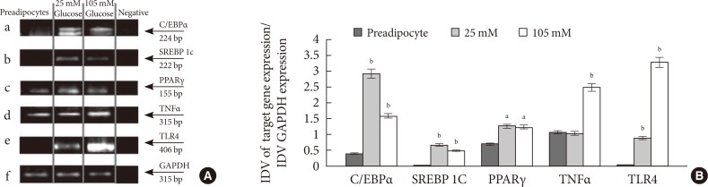
Effect of 105 mM glucose on molecular markers involved in the process of adipogenesis and inflammation at gene level. (A) CCAAT-enhancer-binding proteins α (C/EBPα) (a); sterol regulatory element-binding protein 1c (SREBP 1c) (b); peroxisome proliferator-activated receptor γ (PPARγ) (c); tumor necrosis factor α (TNFα) (d); toll-like receptor 4 (TLR4) (e); and glyceraldehyde 3-phosphate dehydrogenase (GAPDH) (f). Lane 1 to 4 indicates preadipocytes, adipocytes differentiated with 25, 105 mM glucose and negative control, respectively. GAPDH was used as internal control for the study. (B) The graph represents the ratio of integrated density value (IDV) data of genes to IDV of GAPDH. The 25 and 105 mM glucose expression levels were compared to preadipocytes. aP<0.001, bP<0.0001.
Effect of high glucose concentration on inflammatory targets and adipocytokines
An increased expression of TNFα and TLR4 were observed (Fig. 1Ad, Ae, and B) in the presence of 105 mM glucose, along with an increased total protein level of nuclear factor-κB (NF-κB) (Fig. 2Aa and B) in comparison to control cells (25 mM glucose). Cells differentiated in the presence of 105 mM glucose exhibited decreased adiponectin expression at translational level (Fig. 2Ab and B).
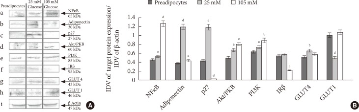
Effect of 105 mM glucose on molecular markers involved in the process of proliferation, adipogenesis, inflammation and insulin signaling at protein level. (A) nuclear factor-κB (NF-κB) (a); Adiponectin (b); p27 (c); Akt/phosphoprotein kinase B (PKB) (d); phosphoinositide 3 kinase (PI3K) (e); insulin receptor β (IRβ) (f); glucose transporter 4 (GLUT4) (g); GLUT1 (h); and β-actin (i). Lane 1, 2, and 3 indicate preadipocytes, adipocytes differentiated with 25 and 105 mM glucose, respectively. β-Actin was used as internal control for the study. (B) The graph represents the ratio of integrated density value (IDV) data of proteins to IDV of β-actin. The 25 and 105 mM glucose expression levels were compared to preadipocytes. aP<0.05, bP<0.01, cP<0.001, dP<0.0001.
Effect of high glucose concentration on various cell proliferative markers
p27 expression was observed in cells differentiated in 25 mM glucose, whereas a complete inhibition of protein expression was observed in cells differentiated in 105 mM glucose (Fig. 2Ac and B). An increase in phosphoprotein kinase B (Akt/PKB) and phosphoinositide 3 kinase (PI3K) protein expression was observed in cells differentiated with 105 mM glucose compared to 25 mM glucose (Fig. 2Ad, Ae, and B).
Effect of high glucose concentration on glucose uptake and metabolism
A 37.9%±2.1% (P<0.01) increase in glucose uptake and 25.7%±0.03% (P<0.0001) decrease in glycogen synthesis was observed in cells differentiated with 105 mM glucose in comparison to control (25 mM glucose).
Effect of high glucose concentration on various molecular markers involved in insulin signaling
A decrease in protein expression of insulin receptor β was observed (Fig. 2Af and B) at 105 mM glucose, whereas glucose transporter 4 (GLUT4) levels remained unaffected (Fig. 2Ag and B), suggesting glucose uptake via insulin independent pathway. An increased level of GLUT1 in comparison to control (25 mM glucose) was observed in cells differentiated with 105 mM glucose (Fig. 2Ah and B). β-Actin was used as internal control for protein expression studies (Fig. 2Ai).
DISCUSSION
Han et al. [11] have reported insulin resistance and hypertrophy in 3T3-L1 cells cultured with 25 mM glucose. To probe the role of glucose in obesity, the effect of various concentrations of glucose on 3T3-L1 cells were analyzed. Interestingly, 3T3-L1 cells in 105 mM glucose showed a decrease in adipogenesis compared to 25 mM glucose. MTT assay and [3H]-thymidine incorporation study indicated an increase in proliferation of cells treated with 105 mM glucose, confirming that the decreased lipid accumulation with 105 mM glucose is not due to cytotoxicity. The decrease in the expression of adipogenic targets, indicating a decrease in adipogenesis, could be due to the continuous presence of high glucose, causing stress, and leading to an inhibition of differentiation of preadipocytes into functional adipocytes [4-6]. Inflammatory stimuli trigger the activation of TLRs and the release of proinflammatory cytokines leading to increased transcription of NF-κB target genes [12]. Overexpression of NF-κB activates cellular stress signals and inflammatory cytokines, including TNFα, thereby disturbing the cellular signaling. Increased secretion of TNFα has been known to inhibit differentiation of preadipocytes into functional adipocytes, with its elevated level accounting for an increased population of activated preadipocytes [6] and decreased production of adiponectin, which are implicated in the development of obesity [13]. Further investigation of p27 at a concentration of 105 mM glucose validated the induction of hyperplasia in 3T3-L1 cells [14,15]. The increase in Akt/PKB and PI3K levels in cells differentiated in 105 mM glucose medium suggested PI3K dependent proliferation of cells. An increase in glucose uptake and decrease in glycogen synthesis was observed in cells differentiated in 105 mM glucose. Thereafter, evaluation of the pathway initiating glucose uptake, showed a decrease in IR expression, with the total GLUT4 levels remaining unaltered, indicating an insulin independent pathway.
In conclusion, the study clearly demonstrated that an uncontrolled proliferation of activated preadipocytes due to prolonged presence of high glucose concentration (105 mM) can lead to the development of adipocyte hyperplasia.
ACKNOWLEDGMENTS
Kusampudi Shilpa (SRF, 142049/2K10/1) is grateful to the Council of Scientific and Industrial Research, Government of India, for financial assistance.
Notes
No potential conflict of interest relevant to this article was reported.