
- Current
- Browse
- Collections
-
For contributors
- For Authors
- Instructions to authors
- Article processing charge
- e-submission
- For Reviewers
- Instructions for reviewers
- How to become a reviewer
- Best reviewers
- For Readers
- Readership
- Subscription
- Permission guidelines
- About
- Editorial policy
Articles
- Page Path
- HOME > Diabetes Metab J > Volume 35(1); 2011 > Article
-
ReviewNew Perspectives on Diabetic Vascular Complications: The Loss of Endogenous Protective Factors Induced by Hyperglycemia
- In-Kyung Jeong1, George L. King2
-
Diabetes & Metabolism Journal 2011;35(1):8-11.
DOI: https://doi.org/10.4093/dmj.2011.35.1.8
Published online: February 28, 2011
1Department of Endocrinology and Metabolism, Kyung Hee University School of Medicine, Seoul, Korea.
2Section on Vascular Cell Biology, Joslin Diabetes Center, Harvard University, Boston, MA, USA.
- Corresponding author: George L. King. Section on Vascular Cell Biology, Joslin Diabetes Center One Joslin Place, Boston, MA 02215, USA. George.King@joslin.harvard.edu
Copyright © 2011 Korean Diabetes Association
This is an Open Access article distributed under the terms of the Creative Commons Attribution Non-Commercial License (http://creativecommons.org/licenses/by-nc/3.0/) which permits unrestricted non-commercial use, distribution, and reproduction in any medium, provided the original work is properly cited.
ABSTRACT
- Diabetic vascular complications are among the leading causes of morbidity and mortality in diabetic patients. In the past, many studies have focused on the mechanisms of hyperglycemia-induced chronic vascular complications via the formation of toxic metabolites such as oxidative stress, advanced glycosylated end products, persistent activation of protein kinase C, and increased sorbitol concentrations. However, vascular complications result from imbalances caused by increases in systemic toxic metabolites, such as those that occur under conditions of hyperglycemia and dyslipidemia, and by reductions in endogenous protective factors such as insulin, vascular endothelial growth factor, and platelet derived growth factor. This review outlines some of the evidence supporting the importance of enhancing endogenous regenerative factors.
- 1. Brownlee M. Biochemistry and molecular cell biology of diabetic complications. Nature 2001;414:813-820. ArticlePubMedPDF
- 2. Enge M, Bjarnegard M, Gerhardt H, Gustafsson E, Kalen M, Asker N, Hammes HP, Shani M, Fassler R, Betsholtz C. Endothelium-specific platelet-derived growth factor-B ablation mimics diabetic retinopathy. EMBO J 2002;21:4307-4316. ArticlePubMedPMC
- 3. Yokota T, Ma RC, Park JY, Isshiki K, Sotiropoulos KB, Rauniyar RK, Bornfeldt KE, King GL. Role of protein kinase C on the expression of platelet-derived growth factor and endothelin-1 in the retina of diabetic rats and cultured retinal capillary pericytes. Diabetes 2003;52:838-845. ArticlePubMedPDF
- 4. Geraldes P, Hiraoka-Yamamoto J, Matsumoto M, Clermont A, Leitges M, Marette A, Aiello LP, Kern TS, King GL. Activation of PKC-delta and SHP-1 by hyperglycemia causes vascular cell apoptosis and diabetic retinopathy. Nat Med 2009;15:1298-1306. ArticlePubMedPMCPDF
- 5. Sison K, Eremina V, Baelde H, Min W, Hirashima M, Fantus IG, Quaggin SE. Glomerular structure and function require paracrine, not autocrine, VEGF-VEGFR-2 signaling. J Am Soc Nephrol 2010;21:1691-1701. ArticlePubMedPMC
- 6. Ziyadeh FN. Different roles for TGF-beta and VEGF in the pathogenesis of the cardinal features of diabetic nephropathy. Diabetes Res Clin Pract 2008;82(Suppl 1):S38-S41. PubMed
- 7. Eremina V, Jefferson JA, Kowalewska J, Hochster H, Haas M, Weisstuch J, Richardson C, Kopp JB, Kabir MG, Backx PH, Gerber HP, Ferrara N, Barisoni L, Alpers CE, Quaggin SE. VEGF inhibition and renal thrombotic microangiopathy. N Engl J Med 2008;358:1129-1136. ArticlePubMedPMC
- 8. He Z, Opland DM, Way KJ, Ueki K, Bodyak N, Kang PM, Izumo S, Kulkarni RN, Wang B, Liao R, Kahn CR, King GL. Regulation of vascular endothelial growth factor expression and vascularization in the myocardium by insulin receptor and PI3K/Akt pathways in insulin resistance and ischemia. Arterioscler Thromb Vasc Biol 2006;26:787-793. ArticlePubMed
- 9. Rask-Madsen C, King GL. Mechanisms of disease: endothelial dysfunction in insulin resistance and diabetes. Nat Clin Pract Endocrinol Metab 2007;3:46-56. ArticlePubMedPDF
- 10. Rask-Madsen C, Li Q, Freund B, Feather D, Abramov R, Wu IH, Chen K, Yamamoto-Hiraoka J, Goldenbogen J, Sotiropoulos KB, Clermont A, Geraldes P, Dall'Osso C, Wagers AJ, Huang PL, Rekhter M, Scalia R, Kahn CR, King GL. Loss of insulin signaling in vascular endothelial cells accelerates atherosclerosis in apolipoprotein E null mice. Cell Metab 2010;11:379-389. ArticlePubMedPMC
- 11. Keenan HA, Costacou T, Sun JK, Doria A, Cavellerano J, Coney J, Orchard TJ, Aiello LP, King GL. Clinical factors associated with resistance to microvascular complications in diabetic patients of extreme disease duration: the 50-year medalist study. Diabetes Care 2007;30:1995-1997. PubMed
REFERENCES
Fig. 1Diabetes induces an imbalance between toxic and protective factors to cause complications. FFA, free fatty acid; AGE, advanced glycosylated end product; ROS, reactive oxygen species; PKC, protein kinase C; PDGF, platelet-derived growth factor; VEGF, vascular endothelial growth factor; APC, activated protein C.
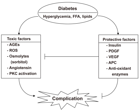
Figure & Data
References
Citations
Citations to this article as recorded by 
- Role of platelet-derived growth factor c on endothelial dysfunction in cardiovascular diseases
Adriana Grismaldo, Luis Sobrevia, Ludis Morales
Biochimica et Biophysica Acta (BBA) - General Subjects.2022; 1866(10): 130188. CrossRef - Ginkgo biloba extracts protect human retinal Müller glial cells from t-BHP induced oxidative damage by activating the AMPK-Nrf2-NQO-1 axis
Yue Li, Ke Wang, Xue Zhu, Zhengqi Cheng, Ling Zhu, Michael Murray, Fanfan Zhou
Journal of Pharmacy and Pharmacology.2022;[Epub] CrossRef - Organic Isothiocyanates as Hydrogen Sulfide Donors
Alma Martelli, Valentina Citi, Lara Testai, Simone Brogi, Vincenzo Calderone
Antioxidants & Redox Signaling.2020; 32(2): 110. CrossRef - PGC‐1α, a potential therapeutic target against kidney aging
Gayoung Lee, Md Jamal Uddin, Yoojeong Kim, Minji Ko, Inyoung Yu, Hunjoo Ha
Aging Cell.2019;[Epub] CrossRef - Glucometabolic characteristics and higher vascular complication risk in Korean patients with type 2 diabetes with non-albumin proteinuria
Yongin Cho, Yong-ho Lee, Eun Seok Kang, Bong-soo Cha, Byung-wan Lee
Journal of Diabetes and its Complications.2019; 33(8): 585. CrossRef - Ameliorative effects of taurine against diabetes: a review
Inam-u-llah, Fengyuan Piao, Rana Muhammad Aadil, Raheel Suleman, Kaixin Li, Mengren Zhang, Pingan Wu, Muhammad Shahbaz, Zulfiqar Ahmed
Amino Acids.2018; 50(5): 487. CrossRef - Notoginsenoside Fc attenuates high glucose-induced vascular endothelial cell injury via upregulation of PPAR-γ in diabetic Sprague–Dawley rats
Jingjing Liu, Chunyu Jiang, Xu Ma, Jianbo Wang
Vascular Pharmacology.2018; 109: 27. CrossRef - Grape seed proanthocyanidin extract protects the retina against early diabetic injury by activating the Nrf2 pathway
YAN SUN, CAIMEI XIU, WEI LIU, YUAN TAO, JIANRONG WANG, YI QU
Experimental and Therapeutic Medicine.2016; 11(4): 1253. CrossRef - Hydrogen peroxide prevents vascular calcification induced ROS production by regulating Nrf-2 pathway
Wensong Zhang, Yi Li, Hanlu Ding, Yaqin Du, Li Wang
Renal Failure.2016; 38(7): 1099. CrossRef - TNFSF15 Inhibits Blood Retinal Barrier Breakdown Induced by Diabetes
Feng Jiang, Qingzhong Chen, Liming Huang, Ying Wang, Zhuhong Zhang, Xiangda Meng, Yuanyuan Liu, Chunjie Mao, Fang Zheng, Jingkai Zhang, Hua Yan
International Journal of Molecular Sciences.2016; 17(5): 615. CrossRef - Effect of Triflusal on Primary Vascular Dysregulation Compared with Aspirin: A Double-Blind, Randomized, Crossover Trial
Sanghoon Shin, Kwang-Joon Kim, In-Jeong Cho, Geu-Ru Hong, Yangsoo Jang, Namsik Chung, Young Min Rah, Hyuk-Jae Chang
Yonsei Medical Journal.2015; 56(5): 1227. CrossRef - NRF2 plays a protective role in diabetic retinopathy in mice
Zhenhua Xu, Yanhong Wei, Junsong Gong, Hongkwan Cho, James K. Park, Ee-Rah Sung, Hu Huang, Lijuan Wu, Charles Eberhart, James T. Handa, Yunpeng Du, Timothy S. Kern, Rajesh Thimmulappa, Alistair J. Barber, Shyam Biswal, Elia J. Duh
Diabetologia.2014; 57(1): 204. CrossRef - Inhibition of JNK by compound C66 prevents pathological changes of the aorta in STZ‐induced diabetes
Yucheng Liu, Yonggang Wang, Xiao Miao, Shanshan Zhou, Yi Tan, Guang Liang, Yang Zheng, Quan Liu, Jian Sun, Lu Cai
Journal of Cellular and Molecular Medicine.2014; 18(6): 1203. CrossRef - Sulforaphane Attenuation of Type 2 Diabetes-Induced Aortic Damage Was Associated with the Upregulation of Nrf2 Expression and Function
Yonggang Wang, Zhiguo Zhang, Wanqing Sun, Yi Tan, Yucheng Liu, Yang Zheng, Quan Liu, Lu Cai, Jian Sun
Oxidative Medicine and Cellular Longevity.2014; 2014: 1. CrossRef - Therapeutic Effect of MG132 on the Aortic Oxidative Damage and Inflammatory Response in OVE26 Type 1 Diabetic Mice
Xiao Miao, Wenpeng Cui, Weixia Sun, Ying Xin, Bo Wang, Yi Tan, Lu Cai, Lining Miao, Yaowen Fu, Guanfang Su, Yuehui Wang
Oxidative Medicine and Cellular Longevity.2013; 2013: 1. CrossRef - Risk Factors for the Progression of Intima-Media Thickness of Carotid Arteries: A 2-Year Follow-Up Study in Patients with Newly Diagnosed Type 2 Diabetes
Sang Ouk Chin, Jin Kyung Hwang, Sang Youl Rhee, Suk Chon, You-Cheol Hwang, Seungjoon Oh, Kyu Jeung Ahn, Ho Yeon Chung, Jeong-taek Woo, Sung-Woon Kim, Young Seol Kim, Ja-Heon Kang, In-Kyung Jeong
Diabetes & Metabolism Journal.2013; 37(5): 365. CrossRef - Downregulation of Nrf2 and HO-1 expression contributes to oxidative stress in type 2 diabetes mellitus: A study in Juana Koslay City, San Luis, Argentina
Susana Siewert, Irma González, Lucas Santillán, Roberto Lucero, Marta S. Ojeda, María S. Gimenez
Journal of Diabetes Mellitus.2013; 03(02): 71. CrossRef - The effect of oral prostaglandin analogue on painful diabetic neuropathy: a double‐blind, randomized, controlled trial
S. Shin, K. J. Kim, H. ‐J. Chang, B. ‐W. Lee, W. ‐I. Yang, B. ‐S. Cha, D. Choi
Diabetes, Obesity and Metabolism.2013; 15(2): 185. CrossRef - Areca nut procyanidins ameliorate streptozocin-induced hyperglycemia by regulating gluconeogenesis
Pei-Ling Huang, Chin-Wen Chi, Tsung-Yun Liu
Food and Chemical Toxicology.2013; 55: 137. CrossRef - Serum glycated albumin predicts the progression of carotid arterial atherosclerosis
Sun Ok Song, Kwang Joon Kim, Byung-Wan Lee, Eun Seok Kang, Bong Soo Cha, Hyun Chul Lee
Atherosclerosis.2012; 225(2): 450. CrossRef - Sulforaphane prevention of diabetes-induced aortic damage was associated with the up-regulation of Nrf2 and its down-stream antioxidants
Xiao Miao, Yang Bai, Weixia Sun, Wenpeng Cui, Ying Xin, Yuehui Wang, Yi Tan, Lining Miao, Yaowen Fu, Guanfang Su, Lu Cai
Nutrition & Metabolism.2012;[Epub] CrossRef - Association between EPCs count and rate of coronary revascularization in asymptomatic type 2 diabetic patients
Hyun Min Kim, Kwang Joon Kim, Jae Hoon Moon, Hye-Jeong Lee, Min Kyung Chae, Hyuk-Jae Chang, Eun Seok Kang, Bong Soo Cha, Hyun Chul Lee, Young Jin Kim, Byung-Wan Lee
Acta Diabetologica.2012; 49(6): 413. CrossRef - Taurine ameliorates hyperglycemia and dyslipidemia by reducing insulin resistance and leptin level in Otsuka Long-Evans Tokushima fatty (OLETF) rats with long-term diabetes
Kyoung Soo Kim, Da Hee Oh, Jung Yeon Kim, Bong Gn Lee, Jeong Soon You, Kyung Ja Chang, Hyunju Chung, Myung Chul Yoo, Hyung-In Yang, Ja-Heon Kang, Yoo Chul Hwang, Kue Jeong Ahn, Ho-Yeon Chung, In-Kyung Jeong
Experimental & Molecular Medicine.2012; 44(11): 665. CrossRef - Inhibition of NF-κB prevents high glucose-induced proliferation and plasminogen activator inhibitor-1 expression in vascular smooth muscle cells
In-Kyung Jeong, Da Hee Oh, Seung-Joon Park, Ja-Heon Kang, Sunshin Kim, Myung-Shik Lee, Myung-Jun Kim, Yoo-Chul Hwang, Kyu Jeong Ahn, Ho-Yeon Chung, Min-Kyung Chae, Hyung-Joon Yoo
Experimental and Molecular Medicine.2011; 43(12): 684. CrossRef
 PubReader
 Cite
Cite


